
Saltar al contenido
Volver a Labs
BioLab · Casos de investigación

Case Study: Catalina

Ovarian cancerA longitudinal evidence-based case study

Understanding the clinical course, treatment context and evolving evidence.

  • Revisión pública 4 ·

Referencia educativa · No es consejo médico individualizado.

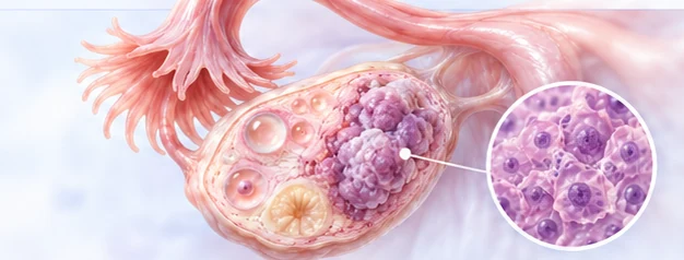
Educational conceptual illustration of an ovary with a magnified inset of cells. It is not a clinical image of Catalina and is not evidence of tumor appearance, subtype, extent or stage.
Conceptual illustration

Línea de tiempo clínica

Los eventos se irán llenando a partir de registros revisados.

Volver a la línea de tiempo

Estudio

Diaphragmatic dome biopsy reported compatible with papillary serous cystadenocarcinoma of the ovary

Fecha del evento
2020-10-21 Fecha exacta
Fecha del documento
2020-10-21
Fecha de registro
2026-11-05
Tipo de evidencia
Informe de laboratorio
Referencia de la fuente
Informe de laboratorio con fecha 2020-10-21
Certeza
Documentado
Revisión
Revisión editorial · Editorial review only: transcription from the original report checked against the document (identifiers omitted); publication approved by the case owner on 5 November 2026. No clinical-specialist review. · 2026-11-05

Resumen

A biopsy of the diaphragmatic dome, reported on 21 October 2020 by a private pathologist, showed infiltration by a malignant epithelial neoplasm forming glands and papillae, compatible with papillary serous cystadenocarcinoma of the ovary.

Hallazgos documentados

Irregular fragment 1.4 x 1 x 0.7 cm. Dense fibroconnective tissue with hemorrhagic zones and fibrinous surfaces, infiltrated by a malignant epithelial neoplasm forming glands and dispersed papillae, some with tortuous lumen, lined by atypical hyperchromatic epithelium, without mucin; associated chronic lymphocytic inflammation. Diagnosis: compatible with papillary serous cystadenocarcinoma of the ovary; the report refers to companion reports 730 and 728.

Incertidumbre

The date the biopsy was taken and the procedure are not stated; 21 October 2020 is the date of the report. Reports 730 and 728 were not handed over. 'Compatible with' is the pathologist's wording; no immunohistochemistry is mentioned. The IMSS summary of 2022 records the histology as poorly differentiated adenocarcinoma.

Contexto de tratamiento relacionado

Ningún registro de contexto de tratamiento está vinculado a este evento.

25 eventos

  1. Intervalo

    EstudioDocumentado

    Diagnosis of non-Hodgkin lymphoma

    Non-Hodgkin lymphoma diagnosed in 2000, according to the 2022 medical summary, which gives the year only.

    Tipo de evidencia: Expediente médicoFecha del documento: 2022-05-18

  2. Intervalo

    Administración confirmadaDocumentado

    Radiotherapy for the lymphoma, 30 fractions, completed in 2000

    Radiotherapy for the lymphoma, 30 fractions, completed in 2000 according to the medical summary.

    Tipo de evidencia: Expediente médicoFecha del documento: 2022-05-18

  3. Fecha aproximada

    Desenlace clínicoDocumentado

    Lymphoma surveillance from 2001

    Under surveillance for the lymphoma from 2001, according to the medical summary.

    Tipo de evidencia: Expediente médicoFecha del documento: 2022-05-18

  4. Fecha exacta

    EstudioDocumentado

    Laboratory studies requested, including tumor markers

    On 2 October 2020 a general and digestive surgeon in private practice requested laboratory studies, among them the tumor markers CA-125, carcinoembryonic antigen, CA 19-9, alpha-fetoprotein and human chorionic gonadotropin.

    Tipo de evidencia: Otro documentoFecha del documento: 2020-10-02

  5. Fecha exacta

    EstudioDocumentado

    Lower-abdominal and transvaginal ultrasound requested

    The same request of 2 October 2020 asked for a lower-abdominal and a transvaginal ultrasound, with emphasis on the ovaries, uterus and tubes.

    Tipo de evidencia: Otro documentoFecha del documento: 2020-10-02

  6. Fecha exacta

    EstudioDocumentado

    CT of the abdomen with contrast and plain chest CT requested

    The same request of 2 October 2020 asked for a CT of the abdomen, plain and with oral and intravenous contrast, and a plain chest CT.

    Tipo de evidencia: Otro documentoFecha del documento: 2020-10-02

  7. Fecha exacta

    EstudioDocumentado

    Tumor markers: CA-125 at 531.4 U/mL, flagged outside the reference interval; CEA, CA 19-9, alpha-fetoprotein and beta-hCG within reference

    Blood drawn on 4 October 2020 at a private hospital laboratory (preliminary report printed 5 October) gave a CA-125 of 531.4 U/mL against a reference interval of 0 to 35, flagged by the laboratory as outside the interval; carcinoembryonic antigen, CA 19-9, alpha-fetoprotein and beta-hCG were within their reference intervals.

    Tipo de evidencia: Informe de laboratorioFecha del documento: 2020-10-05

  8. Fecha exacta

    EstudioDocumentado

    General laboratory panel: blood count, coagulation, febrile agglutinins, chemistry, lipids, electrolytes and hormonal profile

    The same preliminary report of 4 October 2020 covered a complete blood count, coagulation times, febrile agglutinins, a 24-element blood chemistry with lipids and electrolytes, and a gynecologic hormonal profile. The laboratory flagged as outside its reference intervals: erythrocytes 5.65 x10^6/uL, mean corpuscular volume 79.3 fL, globulins 3.7 g/dL, lactate dehydrogenase 238 U/L, cholesterol 260 mg/dL, triglycerides 199 mg/dL, total lipids 827 mg/dL and LDL 190 mg/dL. Hemoglobin, platelets, leukocytes, coagulation, kidney and liver values and electrolytes were within reference; febrile agglutinins were negative; the hormonal values were reported next to post-menopausal reference ranges.

    Tipo de evidencia: Informe de laboratorioFecha del documento: 2020-10-05

  9. Fecha exacta

    EstudioDocumentado

    Pelvic ultrasound: ascites; uterus and ovaries without alterations

    A pelvic ultrasound on 5 October 2020 at a private laboratory found abundant free fluid in the pelvis and both iliac fossae (ascites), a uterus of normal sonographic appearance and ovaries without alterations.

    Tipo de evidencia: Informe de imagenFecha del documento: 2020-10-05

  10. Fecha exacta

    EstudioDocumentado

    Transvaginal ultrasound: ascites and endometrial thickening (16 mm); left ovary not evaluable

    A transvaginal ultrasound the same day found abundant diffuse free fluid and a thickened endometrium of 16 mm, to be correlated clinically; the right ovary showed small diffuse follicles and the left ovary could not be evaluated by this method.

    Tipo de evidencia: Informe de imagenFecha del documento: 2020-10-05

  11. Fecha exacta

    EstudioDocumentado

    Abdominal CT with contrast: ascites of undetermined cause and bilateral pleural effusion; no masses or adenopathies reported

    A CT of the abdomen with oral and intravenous contrast, performed on 5 October 2020 for ascites and abdominal pain and reported on 6 October, found an important amount of free fluid in the abdomen and pelvis and a bilateral pleural effusion with left predominance. Liver, spleen, pancreas, adrenals, kidneys, bowel and the uterus were reported normal, with no retroperitoneal, iliac or inguinal adenopathies.

    Tipo de evidencia: Informe de imagenFecha del documento: 2020-10-06

  12. Fecha exacta

    EstudioDocumentado

    Chest CT: bilateral pleural effusion with passive atelectasis, moderate ascites, no suspicious lung lesions

    A chest CT without contrast on 6 October 2020 at a private imaging center found a bilateral pleural effusion, mild on the right and moderate on the left, with passive atelectasis; moderate ascites in the upper abdomen; a 6 mm calcified granuloma in the left upper lobe; and no suspicious lung lesions, lymphadenopathy or bone lesions.

    Tipo de evidencia: Informe de imagenFecha del documento: 2020-10-06

  13. Fecha aproximada

    EstudioDocumentado

    Diagnosis of ovarian cancer, stage IV (pleural effusion), poorly differentiated adenocarcinoma

    Ovarian cancer, stage IV because of a pleural effusion, with a diagnosis of poorly differentiated adenocarcinoma, as stated in the 2022 medical summary.

    Tipo de evidencia: Expediente médicoFecha del documento: 2022-05-18

  14. Fecha exacta

    ProcedimientoDocumentado

    Laparoscopy at another institution

    Laparoscopy performed on 17 October 2020 at an institution other than the IMSS Oncology Hospital, according to the medical summary.

    Tipo de evidencia: Expediente médicoFecha del documento: 2022-05-18

  15. Fecha exacta

    EstudioDocumentado

    Pleural fluid cytology positive for malignancy; pleural biopsy with metastatic adenocarcinoma

    Pleural fluid and a fragment of pleura taken on 17 October 2020 were reported on 22 October 2020 by a private pathologist: the cytology was positive for malignancy and the pleural biopsy showed metastatic infiltration by adenocarcinoma with zones of mucinous aspect.

    Tipo de evidencia: Informe de laboratorioFecha del documento: 2020-10-22

  16. Fecha exacta

    EstudioDocumentado

    Diaphragmatic dome biopsy reported compatible with papillary serous cystadenocarcinoma of the ovary

    A biopsy of the diaphragmatic dome, reported on 21 October 2020 by a private pathologist, showed infiltration by a malignant epithelial neoplasm forming glands and papillae, compatible with papillary serous cystadenocarcinoma of the ovary.

    Tipo de evidencia: Informe de laboratorioFecha del documento: 2020-10-21

  17. Fecha exacta

    Administración confirmadaDocumentado

    Adjuvant chemotherapy, four cycles, completed

    Adjuvant chemotherapy, four cycles, administered at the IMSS Oncology Hospital and completed on 10 March 2021, according to the medical summary.

    Tipo de evidencia: Expediente médicoFecha del documento: 2022-05-18

  18. Fecha exacta

    ProcedimientoDocumentado

    Interval cytoreductive surgery, reported as optimal

    Interval cytoreductive surgery on 17 May 2021, reported as optimal: hysterectomy, bilateral salpingo-oophorectomy, omentectomy and bilateral pelvic lymphadenectomy, without complications.

    Tipo de evidencia: Expediente médicoFecha del documento: 2022-05-18

  19. Fecha exacta

    Desenlace clínicoDocumentado

    Hospital discharge after the cytoreduction

    Discharged on 19 May 2021 after the cytoreduction, without complications, according to the medical summary.

    Tipo de evidencia: Expediente médicoFecha del documento: 2022-05-18

  20. Fecha exacta

    EstudioDocumentado

    Tumor marker reported negative at the gynecologic-oncology evaluation

    At the gynecologic-oncology evaluation of 26 January 2022 the tumor marker was reported negative, according to the medical summary.

    Tipo de evidencia: Expediente médicoFecha del documento: 2022-05-18

  21. Fecha exacta

    Desenlace clínicoDocumentado

    Evaluation of 26 January 2022: stable, no evidence of recurrence, ECOG 0

    At the last gynecologic-oncology evaluation recorded in the summary (26 January 2022) she reported no symptoms, was considered stable with no evidence of recurrence, and had an ECOG performance status of 0.

    Tipo de evidencia: Expediente médicoFecha del documento: 2022-05-18

  22. Fecha exacta

    Desenlace clínicoDocumentado

    No evidence of disease, stable, as of the medical summary

    As of the medical summary of 18 May 2022, the patient was reported without evidence of disease and stable.

    Tipo de evidencia: Expediente médicoFecha del documento: 2022-05-18

  23. Fecha exacta

    Desenlace clínicoDocumentado

    Death at home on 7 May 2026; the certificate records a hydro-electrolytic imbalance due to ovarian cancer

    The patient died at home in Mexico City on 7 May 2026, with medical attention during the illness, according to the death certificate certified that day by a physician. The certificate records the cause of death as a hydro-electrolytic imbalance (interval of about one day) due to ovarian cancer (interval of about six years), and lists non-Hodgkin lymphoma (about 26 years) as another significant condition that did not lead to the death. The death is recorded as due to illness, not accidental or violent; no necropsy was performed.

    Tipo de evidencia: Otro documentoFecha del documento: 2026-05-07

  24. about four months after 26 January 2022, with a control CT (exact date not stated) Fecha desconocida

    Cita programadaDocumentado

    Follow-up appointment with a control CT scan

    A follow-up visit was scheduled about four months after the 26 January 2022 evaluation, with a control CT scan, according to the medical summary.

    Tipo de evidencia: Expediente médicoFecha del documento: 2022-05-18

  25. date not stated; the family associates it with the studies requested on 2 October 2020 Fecha desconocida

    EstudioRecuerdo retrospectivo de la familiaProbable

    The physician's explanation of the diagnosis, as the family recalls it

    As the family recalls the physician's explanation: an ovarian cancer that was hard to localize, found in the uterus rather than in the ovaries, producing ascites whose fluid was already causing a pleural effusion. Surgery to remove the ovaries and the surrounding tissue was suggested, with a biopsy after the operation.

    Tipo de evidencia: Recuerdo de la familiaFecha del documento: Sin fecha de documento

Abrir evento

Researcher sign-in

Inicia sesión con una cuenta de Stackfire que tenga el permiso de casos de investigación.

KBot · Guía de investigación

Respuestas preescritas · Evidencia enlazada

KBot

Hola, soy KBot, la guía de investigación de BioLab. Puedo explicar los casos de investigación, el Literature Workspace, el grafo de evidencia y las herramientas de análisis, y dónde termina cada uno. Elige un tema abajo. Son explicaciones preescritas, no una consulta de IA en vivo.

Información de investigación, no consejo médico individual. No se envía ni se guarda ningún mensaje.